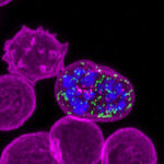
Marapana DS, Lopaticki S, Balan B, Reaksudsan K, Cobbold SA, Geoghegan ND, Khurana S, Scally SW, Hickey P, Jayakrishnan N, Vaibhav V, Spall S, Yousef JM, Dagley LF, Goodman CD, Cozijnsen A, McFadden GI, Rogers KL, Jex A, Komander D, McCarthy JS, Tonkin CJ, Dixon MWA, Cowman AF. GID/CTLH E3 ligase complex control cell fate programs for sexual development of Plasmodium falciparum. Nature Communications. 2026;17(1):10.1038/s41467-026-69183-9
Stanley SE, Carstens RP, Liberti MV, Eertmans W, Vranckx M, Longo D, Ghosal N, Vavrek M, Olsen DB, Cowman AF, Reynders T, Cilissen C, Laethem T, Rottey S, Robbins JA, Stoch SA, Nussbaum J. First-in-human safety and pharmacokinetics of MK-7602, the antimalarial inhibitor of plasmepsins IX/X, in single- and multiple-ascending-dose studies. Antimicrobial Agents and Chemotherapy. 2026;70(2):10.1128/aac.01261-25
Seager BA, Lim PS, Xiao X, Lai KH, Feufack-Donfack LB, Dass S, Jung NC, Abraham A, Grigg MJ, Anstey NM, William T, Sattabongkot J, Leis A, Longley RJ, Duraisingh MT, Popovici J, Wilson DW, Cowman AF, Scally SW. PTRAMP, CSS and Ripr form a conserved complex required for merozoite invasion of Plasmodium species into erythrocytes. Nature Communications. 2026;17(1):10.1038/s41467-026-68486-1
Favuzza P, Palandri J, de Lera Ruiz M, Bailey W, Boyce CW, Danziger A, Fawaz MV, Kelly M, Murgolo N, Robbins JA, Vavrek M, Zhao L, Lei Z, Guo Z, Reaksudsan K, Steel RWJ, Hodder AN, Ngo A, Dziekan JM, Thompson JK, Triglia T, Birkinshaw RW, Penington JS, Scally SW, Dans MG, Coyle R, Sevilleno N, Orban A, Feufack-Donfack LB, Popovici J, Lee MCS, Papenfuss A, Lowes KN, Sleebs BE, McCarthy JS, McCauley JA, Boddey JA, Olsen DB, Cowman AF. MK-7602: a potent multi-stage dual-targeting antimalarial. EBioMedicine. 2026;123:10.1016/j.ebiom.2025.106061
Mansouri M, Dans MG, Low Z, Loi K, Jarman KE, Penington JS, Qiu D, Lehane AM, Crespo B, Gamo F, Baud D, Brand S, Jackson PF, Cowman AF, Sleebs BE. Exploration and Characterization of the Antimalarial Activity of Pyrimidine‐2,4‐Diamines for which Resistance is Mediated by the ABCI3 Transporter. ChemMedChem. 2026;21(1):10.1002/cmdc.202500739
Su W, Nguyen W, Siddiqui G, Dziekan JM, Marapana D, Penington JS, Mehra S, Razook Z, McCann K, Ngo A, Jarman KE, Barry AE, Papenfuss AT, Gilson PR, Creek DJ, Cowman AF, Sleebs BE, Dans MG. Deconvolution of the On-Target Activity of Plasmepsin V Peptidomimetics in Plasmodium falciparum Parasites. ACS Infectious Diseases. 2025;11(12):10.1021/acsinfecdis.5c00742
Kongsomboonvech AK, Scally SW, Le Guen Y, Valissery P, Salinas ND, Cowman AF, Tolia NH, Egan ES. CD44 cross-linking promotes Plasmodium falciparum invasion. Nature Communications. 2025;17(1):10.1038/s41467-025-67030-x
Anton L, Cheng W, Haile MT, Dziekan JM, Cobb DW, Zhu X, Han L, Li E, Nair A, Lee CL, Wang H, Ke H, Zhang G, Doud EH, Cowman AF, Ho C-M. Integrated structural biology of the native malarial translation machinery and its inhibition by an antimalarial drug. Nature Structural & Molecular Biology. 2025;32(11):10.1038/s41594-025-01632-3
Kongsomboonvech AK, Scally SW, Le Guen Y, Valissery P, Salinas ND, Cowman AF, Tolia NH, Egan ES. CD44 cross-linking promotes Plasmodium falciparum invasion. 2025;:10.1101/2025.07.30.667750
Marapana D, Cobbold SA, Pasternak M, Shami GJ, Ralph SA, Lopaticki S, Yousef J, Vaibhav V, Dagley LF, Komander D, Cowman AF. Functional characterisation of components in two Plasmodium falciparum Cullin-RING-Ligase complexes. Scientific Reports. 2025;15(1):10.1038/s41598-025-05342-0