-

Imaging

The latest imaging technologies allow us to explore biological samples like never before. These powerful tools help us ‘unlock visual clues’ to answer the big questions in medical research.

WEHI’s advanced imaging technologies, using optical microscopy combined with powerful computational analysis techniques, are transforming medical research. Our facilities and expertise allow scientists to see spectacular images of biological systems with amazing detail.

These new views of biology, including unprecedented 4D live tissue observations, are bringing us closer to solving some of the world’s most complex health issues.

A game changer for science

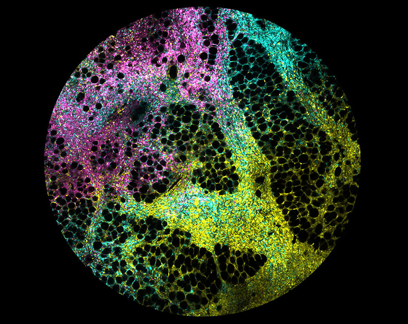
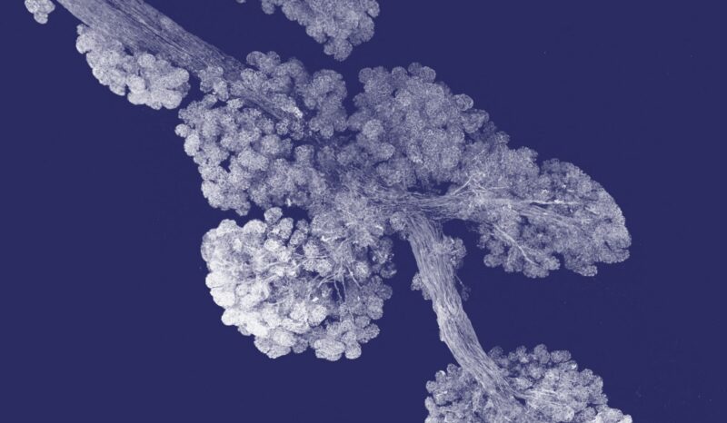
The ability for scientists to visualise biological mechanisms and behaviours is integral to gaining insights into how diseases develop, spread and respond to treatment.

Advanced imaging capabilities, like lattice light sheet microscopy and super resolution microscopy, coupled with advanced bioimage analysis methods, are revealing never-before-seen intricate and informative details of living tissue and systems in 3D and 4D formats.

These insights are sparking a world of new perspectives to accelerate medical discoveries.

“This new technology offers the opportunity to visualise cells and organs in unprecedented detail and in real time. It is a game changer for medical research.”
– Associate Professor Kelly Rogers, Head, Centre for Dynamic Imaging

Imaging at WEHI

WEHI’s imaging technologies support researchers by:

  • creating detailed and data rich images of biological samples
  • enabling analysis and quantification of a range of properties of a sample. For example, cell size and density, cell motility, tumor size and morphology, protein expression and localisation
  • facilitating direct comparison of the effects of drug treatments on various diseases and infections
  • offering a comprehensive set of technologies to image from the single molecule through to the whole organism.

What technologies are at WEHI?

We understand that great science requires state-of-the-art technology.

The Centre for Dynamic Imaging – a research laboratory within WEHI – helps scientists use advanced imaging technology and powerful computational resources to advance research.

Revealing the unseeable – case studies

Advanced imaging technologies are playing an important role in expanding the horizons of medical research.

By capturing and analysing images of living organisms, our scientists are revealing hidden clues to important questions about how our bodies respond to diseases.

These insights are helping us see solutions to problems that have in the past appeared unsolvable.

Light sheet microscopy illuminates brain immune cells


Shown here is a 3D reconstruction of microglia cells (yellow) and blood vessels (blue) in the brain, illuminated using light-sheet microscopy. Visualising the complex network of brain cells helps our researchers further uncover how these cells can influence the success of immunotherapy.

3D reveals inner sanctum of thymus


This 3D reconstruction of the thymus lobe reveals the intricate details of its inner forms. By studying its dynamics, our researchers hope to identify ways to boost thymic function after acute cancer treatments, increasing survival rates for patients.

High-resolution focus on breast cancer

A high-resolution microscope shows this kaleidoscopic image from a tissue fragment within a breast cancer tumour. Each splash of colour represents a unique cancer cell type. This projection enables researchers to better understand how cancer cells move and which cells are more prone to metastasising, helping shine a light on better treatment discoveries. 

Lattice light sheet unlocking malaria secrets


Malaria is one of the deadliest diseases on the planet and affects almost half the world’s population. Understanding the transmission of the disease from human back to mosquitoes could be a major step forward for malaria eradication.

WEHI researchers were able to capture this dynamic transmission in 4D-imaging for the first time, using our world-class lattice light-sheet microscope.

Imaging staff work closely with researchers to design end execute imaging solutions. Access is available to partner research organisations and collaborators.
Celebrating the beauty of life-changing medical research

Art of Science

Art of Science is an annual competition and exhibition of still and moving images captured by WEHI scientists during their research into cancer, infection and immunity and healthy development and ageing.

Sign up to our quarterly newsletter Illuminate

Find out about recent discoveries, community supporters and more.

Illuminate Autumn 2024
View the current issue