-

Centre for Dynamic Imaging

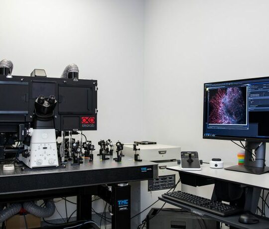
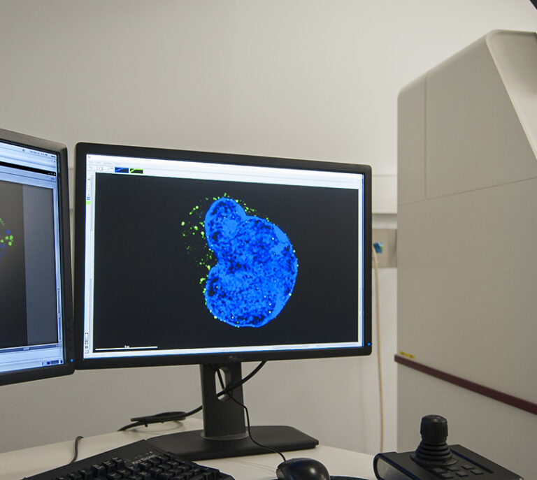
Nikon N-STORM 5.0

A super-resolution fluorescence microscope that localises single molecules in three dimensions to achieve resolutions roughly ten times better than conventional techniques.

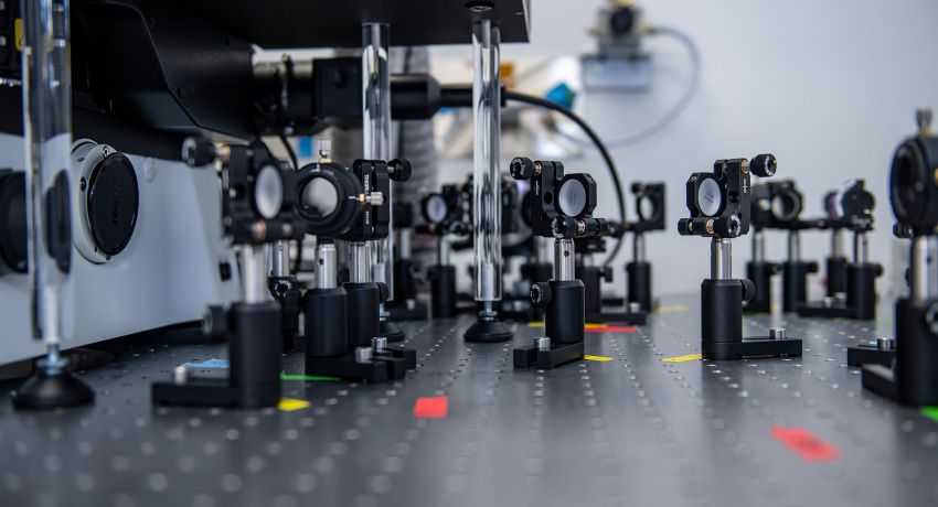
The Nikon N-STORM is a single molecule localisation microscope that is capable of imaging in three-dimensions (3D) with resolutions approximately ten times better than conventional microscopy, typically ~20 nm in xy and ~50 nm in z. The system contains Mic-AO, an adaptive optics module to remove optical aberrations induced by thick samples.

The microscope is also capable of multi-colour, total internal reflection fluorescence (TIRF) and live cell imaging.

The system is compatible with standard slides, 35 mm dishes and 8-well chamber slides. It contains all necessary accessories for environmental control; CO2, humidity, and temperature control.

Staff at the Centre for Dynamic Imaging can provide expert guidance on the preparation of samples and choice of fluorophores.

Objectives

Light sources

  • 405 nm, 488 nm, 568, 640 nm lasers

Cameras

  • sCMOS cameras (Hamamatsu Orca-Flash4.0 V3)
Magnification100x100x
NA1.491.45
Immersion mediaOilSilicone Oil
Working distance (mm)0.120.13

Technological specifications (capabilities)

Available modalities

  • 3D Single Molecule Localization (SML), (also known as PALM, FPALM, STORM, dSTORM, etc)
  • 2D SML
  • TIRF SML
  • Multi-colour SML
  • Z-stacks for thick imaging
  • Fixed cell imaging
  • Live cell imaging
  • Tissue imaging
  • Perfect Focus for drift-free imaging

Unique features

  • The N-STORM is a stable microscope platform, which enables super-resolution imaging in cells and tissues.
  • The Mic-AO adaptive optics system removes optical aberrations to achieve more consistent and uniform resolution throughout thick samples.
  • The system enables multi-colour, 3D imaging.
  • A 100x/1.45 Silicone Oil objective is also available. Silicone Oil has a refractive index very close to biological specimens.

Limitations

  • Ten to hundreds of thousands of camera frames need to be captured to create one super resolved image.
  • Weak staining, excessive bleaching or high levels of background can impair localisation.

Resource

  • Staff in the Centre for Dynamic Imaging can provide documentation to assist with the preparation of samples.

Related microscope

Expert

Dr Michael Mlodzianoski
Super-resolution specialist
Contact us

Contact the Centre for Dynamic Imaging to book equipment or discuss imaging collaborations.

Centre for Dynamic Imaging

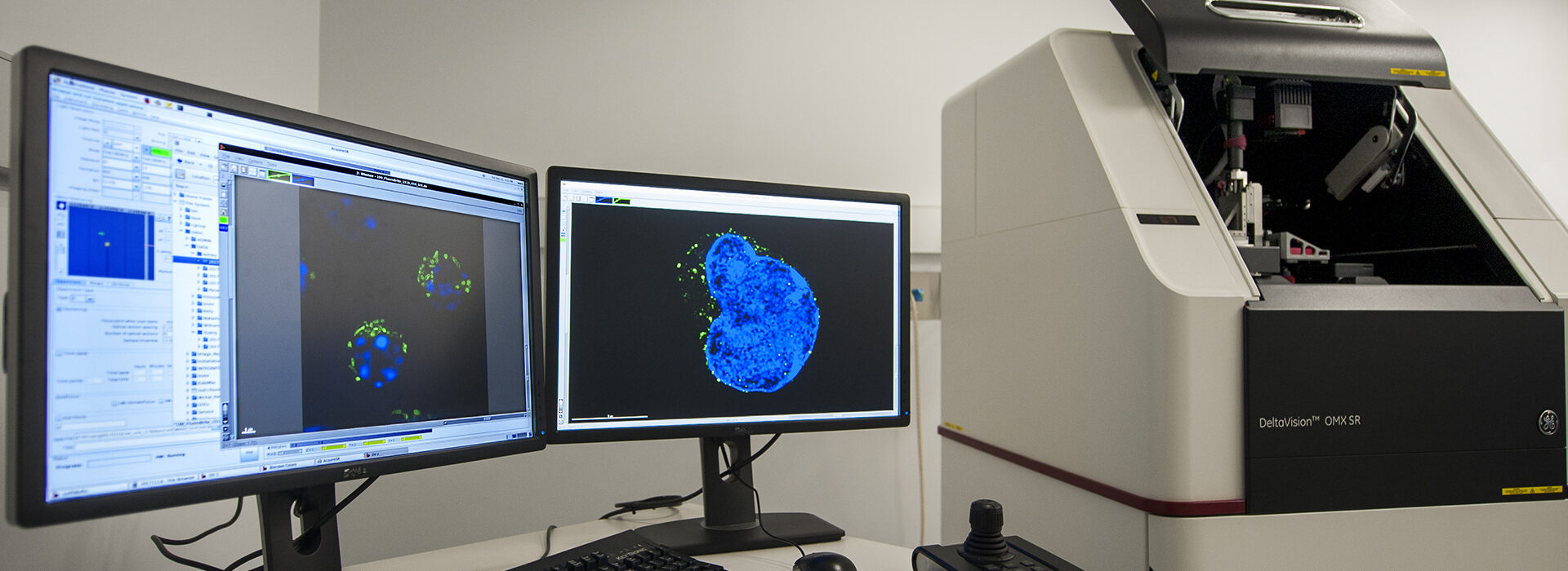
DeltaVision OMX SR

The OMX SR is a microscope for imaging live cells using structured illumination microscopy (SIM). The microscope can rapidly capture very high resolution multicolour images in three dimensions.

The OMX SR is a live cell three-dimensional (3D) structured illumination, super-resolution microscope (3D-SIM).

The system is unique because it enables up to three colours to be captured with two-fold better resolution than conventional microscopy and with high speeds compared to other super-resolution microscopy techniques. The platform also enables two-dimensional SIM and TIRF-SIM (total internal reflection fluorescence structured illumination microscopy). It is also suitable for fixed imaging.

This technique works best on cells or thin specimens (< 15 microns), that are brightly stained.

The system is compatible with standard slides, 35 mm dishes and 8-well chamber slides. It contains all necessary accessories for environmental control, for example CO2, humidity, and temperature control.

Staff at the Centre for Dynamic Imaging can provide expert guidance on the preparation of samples and choice of fluorophores.
Hardware

Light sources

  • 405 nm, 488 nm, 568, 640 nm
  • LED trans-illuminator

Cameras

  • 3 x PCO.edge sCMOS cameras
Objective magnification5x10x20x25x25x
Immersion mediaAirMultiWaterWaterClarity
XY-resolution (µm)4.972.491.240.990.99
Working distance (mm)208228
NA0.100.601.001.051.00

Technological specifications (capabilities)

Available modalities

  • 3D SIM (structural illumination microscopy)
  • 2D SIM
  • TIRF-SIM
  • Very fast and multi-colour simultaneous widefield deconvolution
  • Z-stack
  • Time-series
  • Tile scan
  • Multi positions
  • Live cell imaging

Additional hardware

  • Blaze SIM Module OPTION for DeltaVision OMX SR
  • Ultimate Focus Hardware Autofocus Module
  • DIC optics

Unique features

  • The OMX SR is a stable and compact microscope platform, which enables the dynamic capture of super-resolved images from live cells.
  • The system enables fast three-colour live cell 3D-SIM imaging.
  • A 60x/1.3 Silicone Oil objective is also available for live cell widefield deconvolution microscopy. Silicone Oil has a refractive index more similar to water and remains constant during long-term observations.
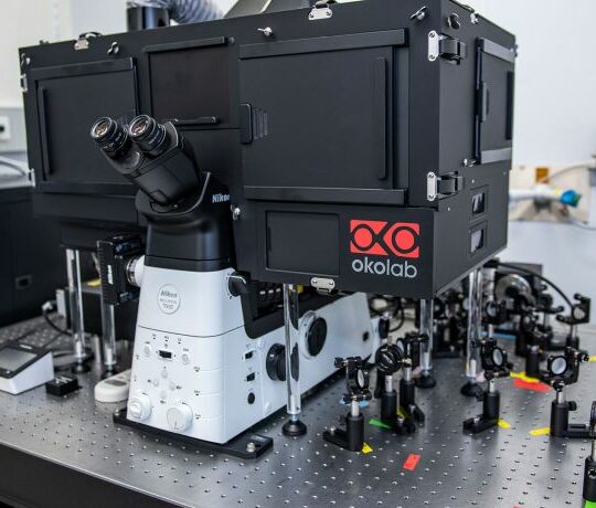
Deltavision OMX microscope at WEHI

Limitations

  • 9 x 15 images need to be captured at every focal plane for 3D-SIM, which means that photobleaching can be difficult.
  • Weak staining, excessive bleaching or high levels of background can cause reconstruction artefacts.

Resource

Staff in the Centre for Dynamic Imaging can provide documentation to assist with the preparation of samples.

Expert

Dr Michael Mlodzianoski
  • Super-resolution specialist
Contact us

Contact the Centre for Dynamic Imaging to book equipment or discuss imaging collaborations.