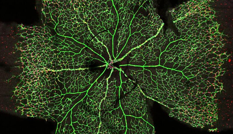
Visualising biological mechanisms and behaviours is a gateway to understanding disease.
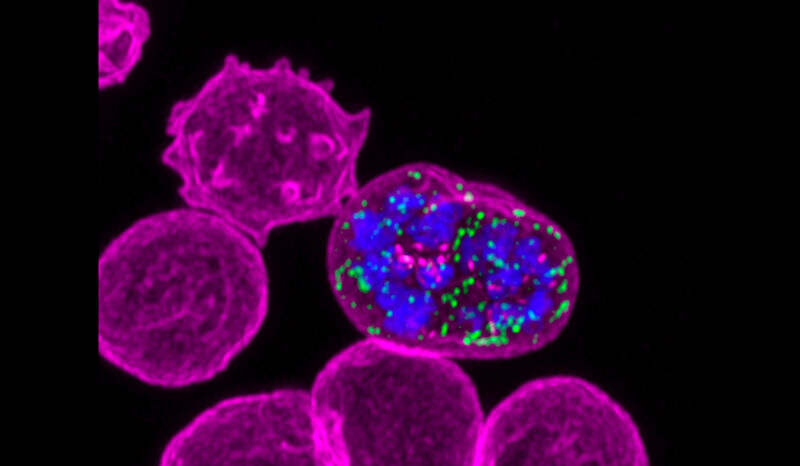
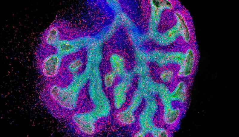
Scientists are now capturing spectacular images and real-time video from single cells and tissue slices, right through to whole organs. These new views of biology are bringing us closer to developing better disease diagnostics and treatments.
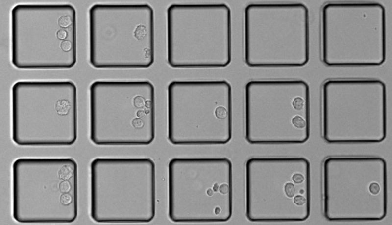
Single cell imaging techniques are allowing researchers, including Dr Kim Pham, to develop complex mathematical models to understand how cancer cells respond to chemotherapy.
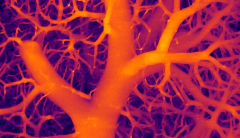
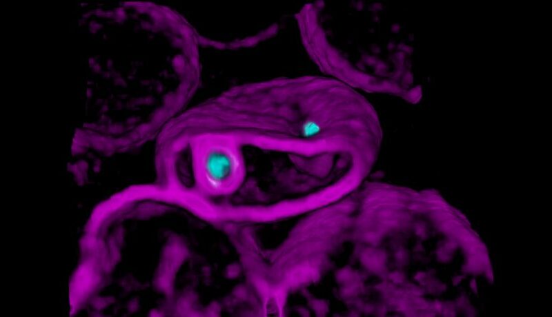
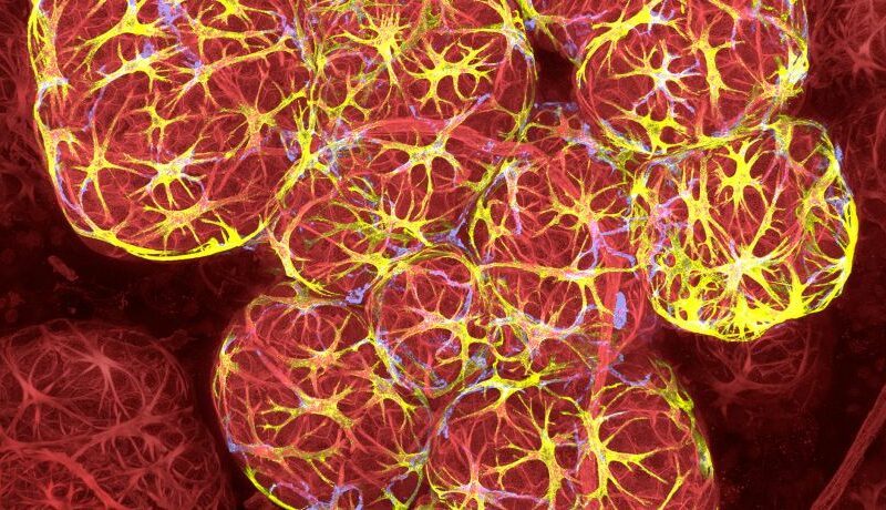
Researchers are using multiplexed, 3-dimensional imaging to combine information about cancer cell movement and blood vessel structure to better understand how tumour cells invade other tissues.
Advanced live-microscopy techniques have become the most effective avenue for studying the interactions between the malaria parasite and the host red blood cell prior to, during, and after infection.
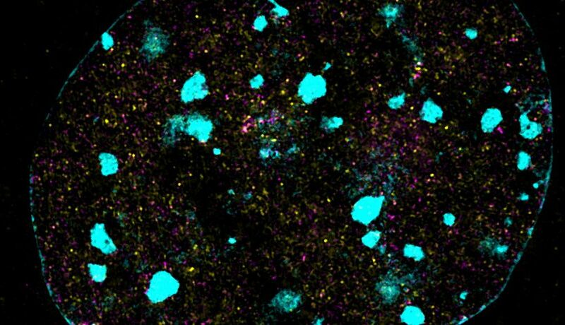
Advanced microscopy techniques are helping researchers like Iromi Wanigasuriya understand how genes are switched on and off, a process called epigenetic control of gene expression.
Access to the Centre’s suite of imaging technologies has enabled WEHI researcher Dr Andre Samson to discover how the killer protein MLKL exerts its deadly job.
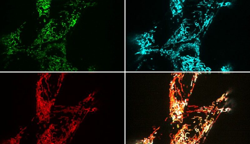
Dr Kate McArthur is using cutting edge imaging techniques to understand how DNA is released from mitochondria, a process that has been implicated in the development of autoimmune disease.