-

Centre for Dynamic Imaging

About us

The Centre for Dynamic Imaging is a research laboratory within WEHI.

We partner with scientists at the institute and across Australia using imaging technology to advance the study of cancer, immune disorders and infectious disease.

Collaborating to improve health outcomes

The centre is run by an interdisciplinary research team with expertise in biomedical engineering, physics, mathematics and biology.

The team builds and enhances imaging equipment, maintaining a combination of bespoke and commercial instruments. The centre also has a strong focus on educating researchers to independently design, apply and analyse their own imaging experiments. This focus has helped researchers use imaging to accelerate scientific discoveries.

The centre receives strategic guidance from a leadership team with expertise in microscopy, research, information technology and business operations.

Philanthropic support is enabling discoveries

The Centre for Dynamic Imaging is supported by a generous gift from the Alan G L Shaw bequest. The donation allowed expansion of the centre, helping to establish it as a world-class imaging facility.

Meet our experts

Professor Kelly Rogers

Professor Kelly Rogers

  • Division Head, Laboratory Head, Centre for Dynamic Imaging Manager

Professor Kelly Rogers has more than 20 years’ experience in advanced microscopy. Her strategic development of the Centre for Dynamic Imaging has enabled researchers to work at the cutting edge of discoveries in human biology.

Areas of expertise:

  • Confocal microscopy
  • Ca2+ imaging
  • Lattice light sheet microscopy
  • Super-resolution microscopy – 3D SIM
  • Preclinical imaging – fluorescence, bioluminescence and micro-CT

Full profile

Dr Niall Geoghegan

  • Lattice light sheet specialist

With a background in engineering and biology Dr Niall Geoghegan is responsible for developing WEHI’s lattice light sheet imaging capabilities.

Areas of expertise:

  • Optical system design and engineering
  • Lattice light sheet microscopy
  • Confocal and widefield technology
  • Time-resolved fluorescence methods: FCS, FLIM, FRAP
  • Live cell imaging

Full profile

Dr Anna Harutyunyan

  • Research Officer

Areas of expertise

  • 3D/Confocal Microscopy
  • Laser microdissection
  • In vivo multiphoton and Ca2+ imaging
  • Multi omics
  • Neurodegenerative diseases

Dr Anna Harutyunyan joined the Centre for Dynamic Imaging in 2024 after completing her PhD in Computational Neuroscience. Prior to her PhD, Anna was classically trained in the Liberal Arts, graduating as Phi Beta Kappa with BSc degrees in Inorganic Chemistry and Molecular Biology. She completed her honours internship at Hershey Medical Center (Penn State University College of Medicine) before joining Carlos Lois’s group at California Institute of Technology (Caltech). There, she employed live Ca2+ imaging and optogenetics in mouse and zebra finch models to study neuronal circuits involved in learning and memory. This work laid the foundation for her deep interest in advanced imaging techniques and neurobiological research.

In 2019, Anna moved to Australia to pursue her PhD at the University of Melbourne. Her doctoral research employed high throughput multi-omics, 3D imaging, and in vivo electrophysiology to investigate the pathological mechanisms underlying Alzheimer’s disease and acquired epilepsy.

At the Centre for Dynamic Imaging, Anna is part of the spatial omics team supporting the MERSCOPE and Multiplex Ion Beam Imaging (MIBIscope) platforms. She also provides support and expertise for experimental design and sample preparation for confocal and in vivo imaging and laser capture microdissection.

Full profile

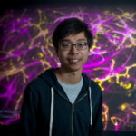
Head-and-shoulders photo of Dawson Ling against a colourful background

Dawson Ling

  • Research Officer

Areas of expertise:

  • Blood-stage malaria biology
  • Live-cell imaging
  • Lattice light sheet microscopy
  • Molecular biology
  • Drug screening

Dawson Ling’s key research interest lies in unravelling how the malaria parasite (Plasmodium falciparum) invades the human red blood cell. He joined the Centre for Dynamic Imaging group to continue working on blood-stage malaria invasion using state-of-the-art microscopy techniques, including lattice light sheet microscopy. He is developing novel imaging techniques, such as expansion microscopy of invading malaria parasites.

He completed his PhD and Honours at the Burnet Institute, where he investigated the mechanism of action of novel invasion-blocking compounds in blood-stage Plasmodium falciparum using a combination of approaches, including functional assays, molecular biology, mass spectrometry and live-cell imaging. During his PhD, he managed to uncover parasite proteins that are involved in invasion-related lipid metabolism/signalling. Previously, he supported clinical research trials as a Clinical Trials Assistant at the Melbourne Children’s Trials Centre (Murdoch’s Children Research Institute) and maintained food quality/assurance as an Analytical Chemist at BVAQ.

Full profile

Dr Claire Marceaux

  • Senior Research Officer

Areas of expertise:

  • Spatial proteomics
  • Multiplex IHC/IF
  • Lung cancer
  • Tumour immune microenvironment
  • Image spatial analysis

Dr Claire Marceaux is a Senior Research Officer at WEHI where she leads spatial proteomics projects in the Center for Dynamic Imaging using the Multiplex Ion Beam Imaging (MIBI) Platform. While managing research assistant and image analyst activities, she develops targeted high-plex panels for multiple type of tissues and the subsequent single-cell image analysis. She is an emerging leader in the field of spatial omics and has been intensively collaborating with academic researchers for novel applications of spatial omics in the medical field.

She drove the analysis of the tumour immune microenvironment of lung cancer using in situ multiplex and high-plex imaging technologies, developing her spatial omics expertise (2019 – 2023, in Marie-Liesse Asselin-Labat’s laboratory). Her work has led to high impact factor publications (Cancer Cell 2023, Cancer Discov, 2020) and collaborations. She was the first user of the MIBIscope in Australia and developed her own panel and pipeline to correlate patient outcomes with lung cancer to their immune tumour infiltration.

Full profile

Dr Pradeep Rajasekhar

  • Bioimage Analyst

Dr Pradeep Rajasekhar is an experienced bioimage analyst who collaborates closely with researchers to implement reproducible workflows.

Full profile

Dr Lachlan Whitehead

  • Bioimage analyst

Dr Lachlan Whitehead has been instrumental in developing the centre’s image analysis capabilities. He works closely with researchers to produce data-rich visualisations of their studies.

Areas of expertise:

  • Image analysis
  • Software
  • 3D design and printing
  • Data visualization
  • Data processing pipelines

Full profile

Dr Raymond Yip

  • Senior Research Officer

Areas of expertise:

  • Spatial transcriptomics
  • Hematological malignancy
  • Breast cancer
  • Bone marrow biology
  • Developmental biology

Dr Raymond Yip is a Senior Research Officer at WEHI with joint appointments across the Imaging, Genomics, and Hawkins laboratories. His research project uses cutting-edge spatial transcriptomics technologies to study how myeloma cancer cells survive and evade therapy inside the bone marrow.

He co-leads WEHI’s spatial transcriptomics team and has extensive collaborations with academic groups and biotechnology companies.

Full profile

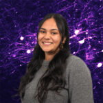
Head-and-shoulders photo of Ishrat Zaman with a purple background

Ishrat Zaman

  • Bioimage Ananalyst

Areas of expertise:

  • Image analysis
  • Bioinformatics
  • Spatial transcriptomics (MERSCOPE)
  • Tissue preparation and imaging
  • Toxoplasma gondii and neuroinflammation

Ishrat Zaman is a bioimage analyst and bioinformatician with experience in both wet-lab and dry-lab environments. She completed her Bachelor of Science with a major in Computational Biology from the University of Melbourne and recently completed her Honours at WEHI, investigating host neuroimmune and neuroinflammatory responses to Toxoplasma gondii infection. Her research involved collecting and analysing spatial transcriptomics, confocal, and Lightsheet data, ranging from 10μm thick sections to whole brain tissues. Currently, she is a member of the Bioimage Analysis Core, where she collaborates with scientists and biologists to analyse their microscopy data. She aims to develop innovative image analysis tools to enhance researchers’ ability to visualise and quantify their data effectively.

Contact us

Contact the Centre for Dynamic Imaging to book equipment or discuss imaging collaborations.