-

Centre for Dynamic Imaging

IVIS Lumina S5

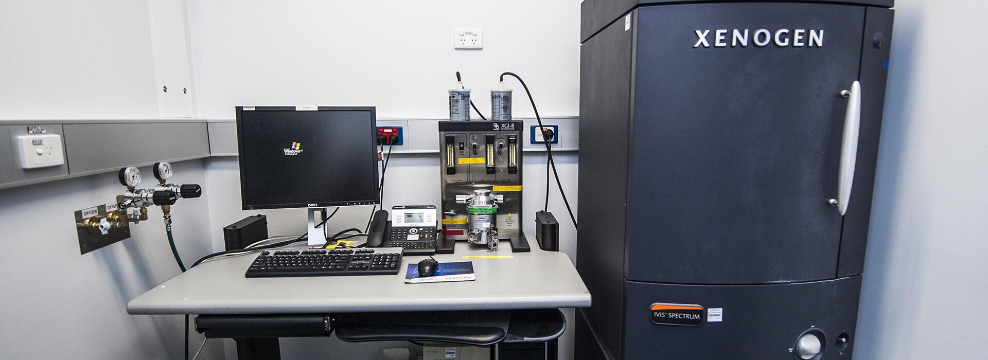
The IVIS Lumina S5 offers scientists the ability to image preclinical models in a non-invasive way in order to monitor disease progression.

The PerkinElmer IVIS Lumina S5 system utilises non-invasive imaging to monitor either bioluminescence or fluorescence located in targeted tumour or diseased cells. This allows for longitudinal imaging experiments to monitor progression of disease states in vivo.

The system allows for a variety of imaging area sizes and can be used with isolation imaging chambers to ensure there is no cross-contamination between experiments. For fluorescent imaging there are 12 excitation filters and 24 emission filters that can be utilised.

A supercooled CCD camera gives users highly sensitive detection with low background noise and includes camera binning options to select for enhanced resolution or enhanced sensitivity depending on the user’s requirements.

Hardware

Detection

16-bit CCD camera

  • Cooled to -90˚C
  • 0-65535 intensity values offers high dynamic range
  • Native resolution of 1920 x 1920 pixels
  • Binning options:
    • Small (4x) – 480 x 480 pixels,
    • Medium (8x) – 240 x 240 pixels; or
    • Large (16x) – 120 x 120 pixels

Imaging area

  • Adjustable field of view: 3.9, 6.5, 13.5 or 21.6 cm2

Light sources

  • 150 W tungsten/halogen lamp for fluorescence

Fluorescence filters

  • 12 excitation filters (430, 465, 500, 535, 570, 605, 640, 675, 710 and 745 nm)
  • 24 emission filters (500, 520, 540, 560, 580, 600, 620, 640, 660, 680, 700, 720, 740, 760, 780, 800, 820 and 840 nm)

Technological specifications (Capabilities)

Available modalities

  • Z-stack
  • Time-series
  • Multi-view (rotation)

Additional options

  • Tile scan possible through use of external macro

Unique features

  • Patterned reference image allows for rudimentary 3D modelling of samples
  • Heated stage ensures specimens are kept warm during imaging procedure

Limitations

  • Fluorescence is not very sensitive
  • Bioluminescent signal fluctuates over time and signal must be of a certain size to be visible
  • Does not allow for single cell resolution or information regarding cellular dynamics

Expert

Professor Kelly Rogers
  • Head, Centre for Dynamic Imaging
Contact us

Contact the Centre for Dynamic Imaging to book equipment or discuss imaging collaborations.

Centre for Dynamic Imaging

Bruker Skyscan 1276 Micro-CT

A high-resolution desktop in vivo micro-CT system with fast InstaRecon® 3D reconstruction software for preclinical imaging.

Micro computed tomography (micro-CT) is X-ray imaging in 3D. The SkyScan 1276 is a high-performance standalone in vivo micro-CT system that gives researchers flexible options for scanning preclinical models and biological samples. Non-destructive handling allows for longitudinal studies of the same sample.

The platform enables both round and spiral (helical) scanning and reconstruction, and low dose imaging for longitudinal studies.

Continuously variable magnification allows users to scan samples with high spatial resolution down to 2.8 μm pixel size.

Variable X-ray energy combined with a range of filters ensures optimal image quality for diverse research applications, including lung tissue and bone.

Hardware

Bruker Skyscan 1276 Micro-CT platform with:

  • 14-bit cooled digital X-ray CCD camera with fibre-optic coupling to scintillator
  • 20-100 kV micro-focus X-ray source
  • 6-positions automatic filter changer for energy selection
  • Integrated physiological monitoring
  • High end computer workstation and InstaRecon® reconstruction software

Technological specifications (capabilities)

  • X-ray source: continuous variable peak energy from 20 till 100kV, < 5 micron spot size at 4 watt; 20W maximum power, air cooled sealed type
  • X-ray detector: 11 Megapixel (4032 x 2688) 14-bit cooled digital X-ray CCD camera with 2×2, 4×4, 8×8 binning modes
  • 6-positions automatic filter changer for energy selection: no filter, Al 0.5mm, Al 1mm, Al 0.5mm + Cu 0.03mm, Cu 0.1mm and “low-dose” filter (beam shaper)
  • Image pixel size can be continuously adjusted from 2.8 μm till 10μm (full camera format) or up to 80μm (8×8 binning)
  • Integrated physiological monitoring (breathing, movement detection, ECG) and real-time motion detection (5 MP colour camera)

Unique features

  • Variable magnification with smallest pixel size of 2.8 micron, 5-6 micron details resolved with more than 10% contrast
  • Spiral scan using endless gantry rotation
  • Step-and-shoot and continuous gantry rotation with shortest scanning cycle of 3.9 sec
  • GPU-accelerated and world’s fastest InstaRecon® 3D reconstructions
  • Integrated physiological monitoring (breathing, movement detection, ECG) for gating and time-resolved 4D micro-tomography
  • Software for 2D/3D image analysis, bone morphology and realistic visualisation
Above: Bone sample imaged on the Bruker Skyscan 1276 Micro-CT. Credit: Raymond Yip
Above: Depth colour coded image of flower. Credit: Lachlan Whitehead

Limitations

  • Resolution is around 6 microns
  • This is not a multi-modality platform
  • Applications requiring lower doses of radiation will have reduced resolution
  • Contrast agents may be required if sample has low X-ray absorption

Related microscope

Experts

Professor Kelly Rogers
  • Head, Centre for Dynamic Imaging
Dr Lachlan Whitehead
  • Bioimage analyst
Contact us

Contact the Centre for Dynamic Imaging to book equipment or discuss imaging collaborations.