-

Centre for Dynamic Imaging

IVIS Lumina S5

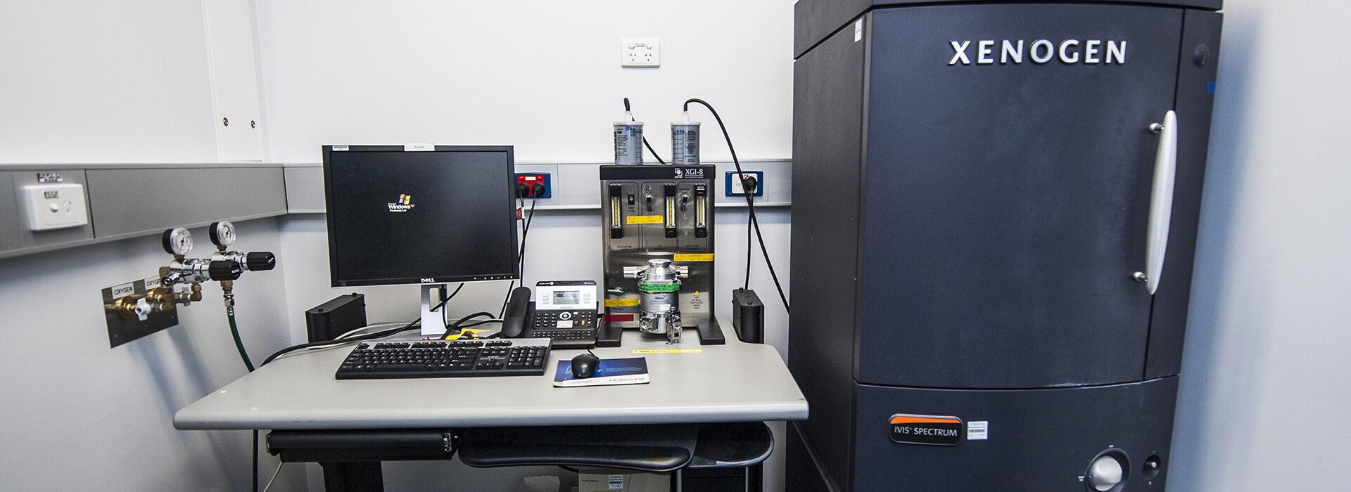
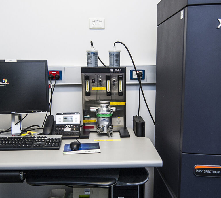
The IVIS Lumina S5 offers scientists the ability to image preclinical models in a non-invasive way in order to monitor disease progression.

The PerkinElmer IVIS Lumina S5 system utilises non-invasive imaging to monitor either bioluminescence or fluorescence located in targeted tumour or diseased cells. This allows for longitudinal imaging experiments to monitor progression of disease states in vivo.

The system allows for a variety of imaging area sizes and can be used with isolation imaging chambers to ensure there is no cross-contamination between experiments. For fluorescent imaging there are 12 excitation filters and 24 emission filters that can be utilised.

A supercooled CCD camera gives users highly sensitive detection with low background noise and includes camera binning options to select for enhanced resolution or enhanced sensitivity depending on the user’s requirements.

Hardware

Detection

16-bit CCD camera

  • Cooled to -90˚C
  • 0-65535 intensity values offers high dynamic range
  • Native resolution of 1920 x 1920 pixels
  • Binning options:
    • Small (4x) – 480 x 480 pixels,
    • Medium (8x) – 240 x 240 pixels; or
    • Large (16x) – 120 x 120 pixels

Imaging area

  • Adjustable field of view: 3.9, 6.5, 13.5 or 21.6 cm2

Light sources

  • 150 W tungsten/halogen lamp for fluorescence

Fluorescence filters

  • 12 excitation filters (430, 465, 500, 535, 570, 605, 640, 675, 710 and 745 nm)
  • 24 emission filters (500, 520, 540, 560, 580, 600, 620, 640, 660, 680, 700, 720, 740, 760, 780, 800, 820 and 840 nm)

Technological specifications (Capabilities)

Available modalities

  • Z-stack
  • Time-series
  • Multi-view (rotation)

Additional options

  • Tile scan possible through use of external macro

Unique features

  • Patterned reference image allows for rudimentary 3D modelling of samples
  • Heated stage ensures specimens are kept warm during imaging procedure

Limitations

  • Fluorescence is not very sensitive
  • Bioluminescent signal fluctuates over time and signal must be of a certain size to be visible
  • Does not allow for single cell resolution or information regarding cellular dynamics

Expert

Professor Kelly Rogers
  • Head, Centre for Dynamic Imaging
Contact us

Contact the Centre for Dynamic Imaging to book equipment or discuss imaging collaborations.