-

Centre for Dynamic Imaging

Zeiss Lightsheet Z.1

The Zeiss Lightsheet Z.1 microscope offers scientists the opportunity to image large 3D objects that would be impossible or far too time consuming to analyse on traditional microscopes.

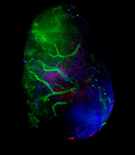
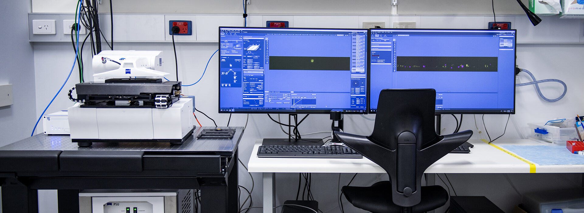
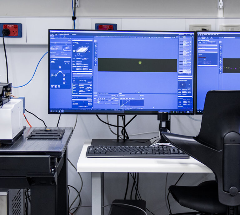
With the Zeiss Z.1 system scientists can utilise a developing technology to acquire large volumetric data sets of 3D structures.

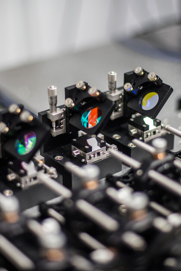
Unlike traditional widefield and confocal microscopes, the Z.1 utilises a ‘sheet’ of light shone through the sample and the image is then detected perpendicular to the lightsheet.

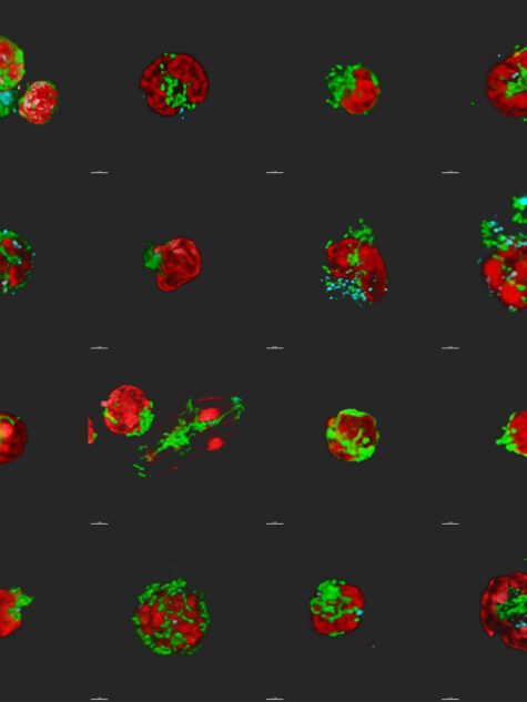
The Z.1 system is intended for fluorescently labelled samples (both endogenous and antibody labelled). It also has the capability of acquiring a bright-field reference image.

The system comes with a number of specific imaging chambers, however sample preparation and mounting are very flexible. This allows for a wide range of sample types to be imaged.

This microscope provides scientists with the ability to image large ‘cleared’ samples to a much greater depth and at faster speeds than has previously been possible.

Hardware

Light sources

  • 405, 445, 488, 514, 561 and 638 nm lasers
  • Bright field LED illumination

Detection source

  • 2 x PCO.Edge sCMOS 16-bit camera
Objective magnification5x20x20x
Immersion mediaAirWaterClarity
XY-resolution (µm)2.220.740.42
Working distance (mm)18.52.45.6
NA0.161.01.0
Above: To image this whole lymph node in 3D, the organ was rendered optically transparent to permit a better penetration of the light for fluorescence microscopy imaging. Credit: Fanny Lafouresse and Verena Wimmer
Above: The Walter and Eliza Hall Institute has a dedicated insectary to assist with development of tools to stop malaria. Credit: Qike Wang and Julie Healer

Technological specifications (capabilities)

Available modalities

  • Z-stack
  • Time-series
  • Multi-view (rotation)

Additional options

  • Tile scan possible through use of external macro

Limitations

  • Resolution limited in Z to lightsheet thickness
  • Tile scan and multi-view unable to be set up together
Above: Auto fluorescence of an ant. Credit: Verena Wimmer
Above: Auto fluorescence of a worm. Credit: Verena Wimmer
Contact us

Contact the Centre for Dynamic Imaging to book equipment or discuss imaging collaborations.

Centre for Dynamic Imaging

Zeiss Lattice Lightsheet (pre-serial instrument)

The Zeiss Lattice Lightsheet is the first pre-commercial system for lattice light sheet microscopy, enabling a greater number of researchers to access the current state of the art technology for live-cell imaging.

Lattice light sheet microscopy is at the cutting edge of technology for live cell imaging due to its combination of high spatial and temporal resolution whilst simultaneously being incredibly gentle on the samples that are imaged.

This technology is now available as a pre-commercial platform offering researchers experimental flexibility in an easy to use system. The technology offers the ability to interrogate complex biological dynamics in 4D with high resolution over long time periods.

Hardware

Objectives

Excitation objective

  • Zeiss 13.3x Magnification, 0.44 NA

Detection Objective

  • Zeiss 44.93x, 1 NA

Imaging characteristics

  • 44.93x Magnification
  • XY Resolution – 330 nm
  • Z resolution – 650 nm
  • Light sheet length – 10 um – 100 µm

Light sources

  • Diode lasers lines – 488, 561 and 640 nm

Detection source

  • PCO.edge sCMOS
  • 2048 x 2048 Pixels
  • 6.5 µm x 6.5 µm pixel size
  • 16 – bit
  • 100 fps (At full frame)
  • 82% QE (Peak at 560 nm)

Technological specifications (Capabilities)

Available modalities

  • Z-stack
  • Time Series
  • Multi-positions

Unique features

  • Compatible with standard sample chambers such as chamber slides, dishes and multi-well plates.
  • Contains transmitted light source for quick and easy sample finding.
  • Can cover a large field of view in a short time.

Limitations

  • Data generated is large and requires post-processing, which takes time

Related microscope

Experts

Cindy Evelyn
Microscopist
Dr Niall Geoghegan
Lattice light sheet specialist
Contact us

Contact the Centre for Dynamic Imaging to book equipment or discuss imaging collaborations.

Centre for Dynamic Imaging

Lattice light sheet

Lattice light sheet microscopy is the current state of the art for live cell imaging, enabling unprecedented 4D imaging capabilities.

Imaging biology in 4D has been a significant challenge in recent decades. The light used for imaging usually damages biological samples, making long term time-lapse microscopy extremely difficult.

Lattice light sheet microscopy is at the cutting edge of technology for live cell imaging because it is incredibly gentle on samples, allowing high resolution imaging over time.

This lattice light sheet was custom-built by staff at the Centre for Dynamic Imaging and was the first of its kind in Australia.

Hardware

Objectives

Excitation objective

  • Special optics 28.6x Magnification, 0.7 NA

Detection Objective

  • Nikon Apo LWD Water dipping objective: 25x, 1.1 NA

Imaging characteristics

  • 62x Magnification
  • XY Resolution – 230 nm
  • Z resolution – 370 nm
  • Light sheet length – 10 µm – 100 µm

Light sources

  • MPB fiber lasers – 488 nm, 561 nm, 589 nm and 642 nm

Detection source

  • 2 x Hamamatsu Orca Flash 4 – V2
  • 2048 x 2048 Pixels
  • 6.5 µm x 6.5 µm pixel size
  • 16 – bit
  • 100 fps (At full frame)
  • 82% QE (Peak at 560 nm)
Above: Immune cell panel: nucleus (red), mitochondria (green) and lysosomes (cyan). Credit: William Chang, Niall Geoghegan
Above: Detailed view of the lattice light sheet microscope

Technological specifications (capabilities)

Available modalities

  • Z-stack
  • Time Series
  • Multi-positions

Unique feature

Lattice light sheet microscopy allows high resolution 4D imaging over many hours.

Limitations

  • Complex sample geometry leads to unconventional sample mounting
  • No transmitted light options
  • No eye pieces
  • Large quantities of data produced

Expert

Dr Niall Geoghegan
  • Lattice light sheet specialist
Contact us

Contact the Centre for Dynamic Imaging to book equipment or discuss imaging collaborations.

Are you requesting a login for the secure online application portal?(Required)
I would like to receive the latest news and updates