-

Centre for Dynamic Imaging

Lunaphore COMET

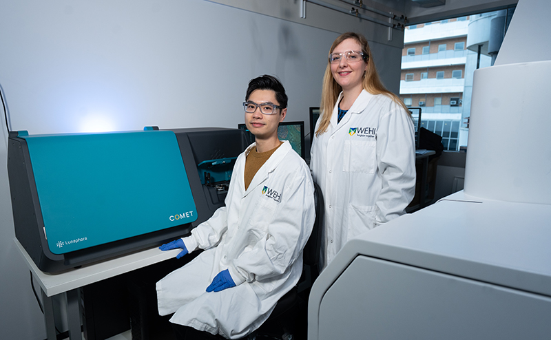
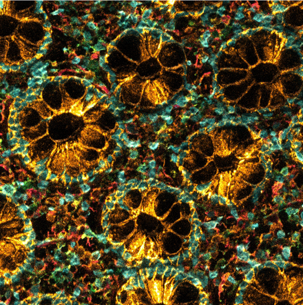
The COMET is a hyperplex spatial biology platform allowing the detection of up to 40 (protein) biomarkers in a single tissue section, preserving spatial context and tissue integrity.

The system uses standard unconjugated, off-the-shelf antibodies to deeply profile the tissue microenvironment, uncover cellular heterogeneity, and accelerate biomarker discovery.

In addition, the COMET allows for the simultaneous detection of up to 12 RNA targets in parallel with protein markers, enabling integrated spatial multiomic analyses in the same sample.

Key experts

Contact us

Contact the Centre for Dynamic Imaging to book equipment or discuss imaging collaborations.

Centre for Dynamic Imaging

Vizgen MERSCOPE

Vizgen MERSCOPE is a high-resolution, in situ spatial genomics platform designed to map and quantify gene expression directly within intact tissue samples at subcellular resolution.

Unique features

It is built around MERFISH (Multiplexed Error-Robust Fluorescence In Situ Hybridization) technology, which allows researchers to design custom gene panels targeting up to 1,000 genes and 6 proteins simultaneously in tissue sections.

Limitations

Users are currently limited to the analysis of 1,000 genes within a user-defined tissue area (up to 1 cm2).

Key experts

Contact us

Contact the Centre for Dynamic Imaging to book equipment or discuss imaging collaborations.

Centre for Dynamic Imaging

Leica LMD7 Laser Microdissection Microscope

Leica LMD7 uses a high-power laser at 349nm to isolate cells and tissue sections mounted on a special membrane slide or dish.

The cutting region of interest (ROI) is pre-defined in the software and can be in any shape. Under the force of gravity, the isolated specimen falls into a collector of choice, such as a PCR tube, the lid of which is attached directly under the specimen holder and is also aligned with the objective that provides the laser beam for cutting as well as other light sources (transmitted light/fluorescence) for sample illumination.

This microdissection approach handles specimens gently without much heat diffusion.

Objectives

MagnificationNumerical Aperture (NA)Working Distance (mm)BF, POL / FL/ DIC, PHLaser CuttingCap InpsectionSpecimen Overview
5x0.1211.7+ / + / –+++++++
10x0.3211.0+ / + / ++++++
20x0.406.9+ / + / ++++++
40x0.603.3-1.9+ / + / ++++
63x0.702.6-1.8+ / + / ++++

BF:brightfield; POL: polarised light; FL: fluorescence; DIC: differential interference contrast; PH: phase contrast.
+: suitable; -: not suitable; ++: dedicated for; +++: most suitable. All are DRY objectives.

Light sources:

  • Brightfield TL: LED (PMax = 15 W) amp housing
  • Fluorescence Lamp Leica EL6000 (120 W metal halide) with filters for DAPI (358nm), GFP (488nm), Y7 (514nm), CY3 (550nm), and CY5 (649nm).
  • Laser Diode-Pumped Solid-State (DPSS), 349 nm (ultraviolet, UV), 120 µJ average energy pulse

Condenser:

  • Condenser head S28, 0.55 NA Motorized 7x condenser disk
  • Motorized polarizer

Detectors:

K3C color camera
K3M monochrome camera

Technical capabilities

Appropriate LMT350 inserts, available are:

  • Multi-collector insert with 4x 0.2mL / 4x 0.5mL PCR tube holders, a single 1.5mL tube holder, and 8/12-well strip tube holders
  • Large slide adaptor
  • 50 mm petri dish adapter
  • 18-well ibidi slide adaptor
  • 96-well PCR / Live Cell Capturing (LCC) plate collector, also compatible to hold 384-well plates but well inspection is limited.

Sample Mounting

  • Appropriate slides or membrane-coated petri dish with samples
  • Glass vs frame
    • High UV absorption. Air exists between membrane and glass backbone. Use of glass slides may require fresh samples for optimal performance
    • Frame slides are great for old samples. Can be challenging for sample mounting but have higher cutting efficiency
    • Usable area
    • Regular slide: usable membrane area (glass) 19 x 41mm, and (frame) 16 x 45mm; total size: 26 x 76mm
    • Large slide: usable membrane area 39 x 45mm; total size: 52 x 76mm
  • PEN slides are versatile, all-purpose membrane slides available in both glass and metal frame formats
  • Choose PEN or PET membrane slides for fixed sample proteomics and metabolomics (free of softeners)
  • PPS membranes have less autofluorescence, a smoother surface but are less resistant to certain chemical treatments
  • Use any type of membrane slides for genomics and transcriptomics
  • Choose DIRECTOR slides to work completely membrane free
  • Keep samples dry as humidity can affect dissected sample collection through gravity
  • 40X and 63X are not available if samples are in the petri dish

Unique Features

The Leica LMD7 can be used to precisely microdissect single cells and regions directly from stained or target-labelled specimen such as H&E and IF tissue sections, preserving molecular integrity for downstream analyses.

This capability enables high-resolution study of archived clinical samples and complex tissue heterogeneity at the single-cell level.

Limitations

A key limitation of laser microdissection is the requirement for samples to be mounted on specialised membrane-coated slides. These membranes are essential to allow precise laser cutting and clean sample collection but add complexity to sample preparation.

Additionally, not all tissue types or staining protocols are compatible with membrane slides, which can limit flexibility and may affect tissue morphology or molecular preservation.

Related microscopes

Key experts

Contact us

Contact the Centre for Dynamic Imaging to book equipment or discuss imaging collaborations.

Centre for Dynamic Imaging

MIBIscope

The MIBIscope is a leading single-cell spatial proteomics technology that acts like a “Google map” for proteins in tissues, revealing where and which proteins are playing a role in both health and disease settings.

The MIBIscope platform uses a cocktail of metal-tagged proteins to map and quantify up to 40 markers within tissues at a single-cell resolution. With the capability to stain simultaneously intact tissues, MIBIscope offers unparalleled versatility in resolving tissue microenvironment and cellular communications across various species and tissue types.

MIBIscope can be used in cancer tissue to study the tumour microenvironment, for example in lung cancer (left).

It can also be used in inflammatory diseases to study immune infiltration, for example in colon tissue (right).

Cancer tissue captured under a microscope and stained in colours

Unique features

Highly multiplexed in situ imaging of proteins. It is compatible with fresh/fixed frozen, and FFPE tissue sections, as well as different species. Human panels for FFPE tissue sections have been validated and are available at the institute.

Customised panels can be developed.

Limitations

It is necessary to put the tissues sections on specific MIBI gold-coated slides.

Resources

Expert

Patrick Lam
  • Microscopist
Michael Mckay
  • Image Bioanalyst
Contact us

Contact the Centre for Dynamic Imaging to book equipment or discuss imaging collaborations.