- About
- Strategic Plan
- Structure
- Governance
- Scientific divisions
- ACRF Cancer Biology and Stem Cells
- ACRF Chemical Biology
- Advanced Technology and Biology
- Bioinformatics
- Blood Cells and Blood Cancer
- Clinical Translation
- Epigenetics and Development
- Immunology
- Infectious Diseases and Immune Defence
- Inflammation
- Personalised Oncology
- Population Health and Immunity
- Structural Biology
- Ubiquitin Signalling
- Laboratory operations
- Funding
- Annual reports
- Human research ethics
- Scientific integrity
- Institute life
- Career opportunities
- Business Development
- Collaborators
- Suppliers
- Publications repository
- Awards
- Discoveries
- Centenary 2015
- History
- Contact us
- Research
- Diseases
- Cancer
- Development and ageing
- Immune health and infection
- Research fields
- Research technologies
- Research centres
- People
- Alistair Brown
- Anne-Laure Puaux
- Assoc Prof Joanna Groom
- Associate Profesor Ian Majewski
- Associate Professor Aaron Jex
- Associate Professor Alyssa Barry
- Associate Professor Andrew Webb
- Associate Professor Chris Tonkin
- Associate Professor Daniel Gray
- Associate Professor Diana Hansen
- Associate Professor Edwin Hawkins
- Associate Professor Ethan Goddard-Borger
- Associate Professor Gemma Kelly
- Associate Professor Grant Dewson
- Associate Professor Isabelle Lucet
- Associate Professor James Vince
- Associate Professor Jason Tye-Din
- Associate Professor Jeanne Tie
- Associate Professor Jeff Babon
- Associate Professor Joan Heath
- Associate Professor John Wentworth
- Associate Professor Justin Boddey
- Associate Professor Kate Sutherland
- Associate Professor Marie-Liesse Asselin-Labat
- Associate Professor Matthew Ritchie
- Associate Professor Melissa Call
- Associate Professor Melissa Davis
- Associate Professor Misty Jenkins
- Associate Professor Nawaf Yassi
- Associate Professor Oliver Sieber
- Associate Professor Peter Czabotar
- Associate Professor Rachel Wong
- Associate Professor Rhys Allan
- Associate Professor Rosie Watson
- Associate Professor Ruth Kluck
- Associate Professor Sandra Nicholson
- Associate Professor Seth Masters
- Associate Professor Sumitra Ananda
- Associate Professor Tim Thomas
- Associate Professor Tracy Putoczki
- Chela Niall
- Deborah Carr
- Dr Alisa Glukhova
- Dr Anna Coussens
- Dr Ashley Ng
- Dr Belinda Phipson
- Dr Ben Tran
- Dr Bernhard Lechtenberg
- Dr Brad Sleebs
- Dr Drew Berry
- Dr Gwo Yaw Ho
- Dr Hamish King
- Dr Hui-Li Wong
- Dr Jacqui Gulbis
- Dr Kelly Rogers
- Dr Lucy Gately
- Dr Margaret Lee
- Dr Mary Ann Anderson
- Dr Maryam Rashidi
- Dr Matthew Call
- Dr Nadia Davidson
- Dr Philippe Bouillet
- Dr Rebecca Feltham
- Dr Rory Bowden
- Dr Samir Taoudi
- Dr Shabih Shakeel
- Dr Shalin Naik
- Dr Sheau Wen Lok
- Dr Stephin Vervoort
- Dr Yunshun Chen
- Guillaume Lessene
- Helene Martin
- Joh Kirby
- Kaye Wycherley
- Keely Bumsted O'Brien
- Mr Mark Eaton
- Mr Simon Monard
- Mr Steve Droste
- Ms Carolyn MacDonald
- Professor Alan Cowman
- Professor Andreas Strasser
- Professor Andrew Lew
- Professor Andrew Roberts
- Professor Anne Voss
- Professor Clare Scott
- Professor David Huang
- Professor David Komander
- Professor David Vaux
- Professor Doug Hilton
- Professor Geoff Lindeman
- Professor Gordon Smyth
- Professor Ian Wicks
- Professor Ivo Mueller
- Professor James McCarthy
- Professor James Murphy
- Professor Jane Visvader
- Professor Jerry Adams
- Professor John Silke
- Professor Ken Shortman
- Professor Leanne Robinson
- Professor Leonard C Harrison
- Professor Lynn Corcoran
- Professor Marc Pellegrini
- Professor Marco Herold
- Professor Marnie Blewitt
- Professor Melanie Bahlo
- Professor Mike Lawrence
- Professor Nicos Nicola
- Professor Peter Colman
- Professor Peter Gibbs
- Professor Phil Hodgkin
- Professor Sant-Rayn Pasricha
- Professor Stephen Nutt
- Professor Suzanne Cory
- Professor Terry Speed
- Professor Tony Papenfuss
- Professor Wai-Hong Tham
- Professor Warren Alexander
- Diseases
- Education
- PhD
- Honours
- Masters
- Clinician-scientist training
- Undergraduate
- Student research projects
- A new regulator of 'stemness' to create dendritic cell factories for immunotherapy
- Advanced imaging interrogation of pathogen induced NETosis
- Cancer driver deserts
- Cryo-electron microscopy of Wnt signalling complexes
- Deciphering the heterogeneity of breast cancer at the epigenetic and genetic levels
- Developing drugs to block malaria transmission
- Developing new computational tools for CRISPR genomics to advance cancer research
- Developing novel antibody-based methods for regulating apoptotic cell death
- Discovering novel paradigms to cure viral and bacterial infections
- Discovery and targeting of novel regulators of transcription
- Dissecting host cell invasion by the diarrhoeal pathogen Cryptosporidium
- Do membrane forces govern assembly of the deadly apoptotic pore?
- Doublecortin-like kinases, drug targets in cancer and neurological disorders
- E3 ubiquitin ligases in neurodegeneration, autoinflammation and cancer
- Engineering improved CAR-T cell therapies
- Epigenetic biomarkers of tuberculosis infection
- Exploiting cell death pathways in regulatory T cells for cancer immunotherapy
- Finding treatments for chromatin disorders of intellectual disability
- Functional epigenomics in human B cells
- Genomic rearrangement detection with third generation sequencing technology
- How does DNA damage shape disease susceptibility over a lifetime?
- How does DNA hypermutation shape the development of solid tumours?
- How platelets prevent neonatal stroke
- Human lung protective immunity to tuberculosis
- Interaction with Toxoplasma parasites and the brain
- Interactions between tumour cells and their microenvironment in non-small cell lung cancer
- Investigating the role of dysregulated Tom40 in neurodegeneration
- Investigating the role of mutant p53 in cancer
- Lupus: proteasome inhibitors and inflammation
- Machine learning methods for somatic genome rearrangement detection
- Malaria: going bananas for sex
- Measurements of malaria parasite and erythrocyte membrane interactions using cutting-edge microscopy
- Measuring susceptibility of cancer cells to BH3-mimetics
- Minimising rheumatic adverse events of checkpoint inhibitor cancer therapy
- Mutational signatures of structural variation
- Naturally acquired immune response to malaria parasites
- Predicting the effect of non-coding structural variants in cancer
- Revealing the epigenetic origins of immune disease
- Reversing antimalarial resistance in human malaria parasites
- Structural and functional analysis of DNA repair complexes
- Targeting human infective coronaviruses using alpaca antibodies
- Towards targeting altered glial biology in high-grade brain cancers
- Uncovering the real impact of persistent malaria infections
- Understanding Plasmodium falciparum invasion of red blood cells
- Understanding how malaria parasites sabotage acquisition of immunity
- Understanding malaria infection dynamics
- Understanding the mechanism of type I cytokine receptor activation
- Unveiling the heterogeneity of small cell lung cancer
- Using alpaca antibodies to understand malaria invasion and transmission
- Using combination immunotherapy to tackle heterogeneous brain tumours
- Using intravital microscopy for immunotherapy against brain tumours
- Using nanobodies to cross the blood brain barrier for drug delivery
- Using structural biology to understand programmed cell death
- School resources
- Frequently asked questions
- Student profiles
- Abebe Fola
- Andrew Baldi
- Anna Gabrielyan
- Bridget Dorizzi
- Casey Ah-Cann
- Catia Pierotti
- Emma Nolan
- Huon Wong
- Jing Deng
- Joy Liu
- Kaiseal Sarson-Lawrence
- Komal Patel
- Lilly Backshell
- Megan Kent
- Naomi Jones
- Rebecca Delconte
- Roberto Bonelli
- Rune Larsen
- Runyu Mao
- Sarah Garner
- Simona Seizova
- Wayne Cawthorne
- Wil Lehmann
- Miles Horton
- Alexandra Gurzau
- Student achievements
- Student association
- Learning Hub
- News
- Donate
- Online donation
- Ways to support
- Support outcomes
- Supporter stories
- Rotarians against breast cancer
- A partnership to improve treatments for cancer patients
- 20 years of cancer research support from the Helpman family
- A generous gift from a cancer survivor
- A gift to support excellence in Australian medical research
- An enduring friendship
- Anonymous donor helps bridge the 'valley of death'
- Renewed support for HIV eradication project
- Searching for solutions to muscular dystrophy
- Supporting research into better treatments for colon cancer
- Taking a single cell focus with the DROP-seq
- WEHI.TV
'Moving target' breast cancer cells revealed by new imaging technique
29 March 2019
Our researchers have developed a new imaging technique to visualise key steps in the evolution of cancer cells within tumours, potentially revealing how breast cancers evade treatment.
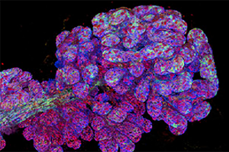
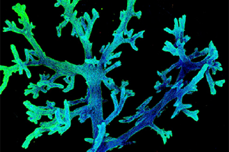
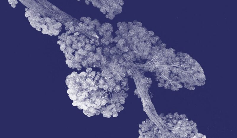
to view tissues and tumours at high resolution that was
previously not possible. This image shows the three-dimensional
structure of mammary ducts of human breast tissue.
Image from Rios, Visvader et al, Cancer Cell.
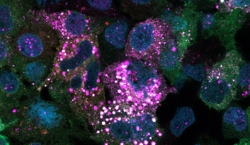
Using a laboratory model of breast cancer, the researchers were able to view tumours in three dimensions, at previously unachievable high resolution. This revealed how cancer cells develop from pre-cancerous cells in the mammary ducts, and changes that occur in the tumour over time.
The research, which was published today in the journal Cancer Cell, suggests that breast cancer cells are inherently changeable, morphing from one cell type to another at the molecular level – resembling cells that are more likely to spread.
At a glance
- A new imaging technique has provided detailed new views of cells in tumours, revealing the changes that occur as breast cancer develops.
- The research revealed that ‘pre-cancerous’ cells in the mammary gland only rarely develop into cancer cells, but that once a cancer forms, the cancer cells appear highly ‘changeable’, a feature that may promote resistance to certain therapies.
Viewing cancer cell evolution
The transformation of normal cells in the mammary gland to cancer cells occurs in many stages, with ‘pre-cancerous’ cells evolving into early-stage cancer cells, which may then undergo changes that make the cells more likely to spread away from the tumour. Until now, it has not been possible to visualise individual clones – ‘sister’ cells that descended from a single pre-cancerous cell – within a whole tumour.
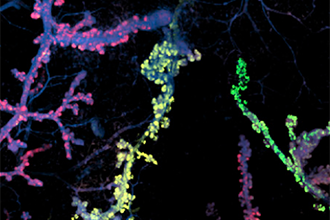
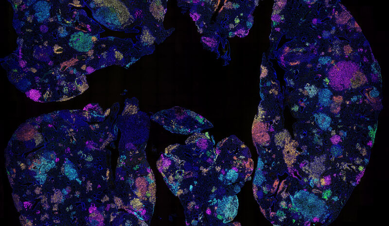
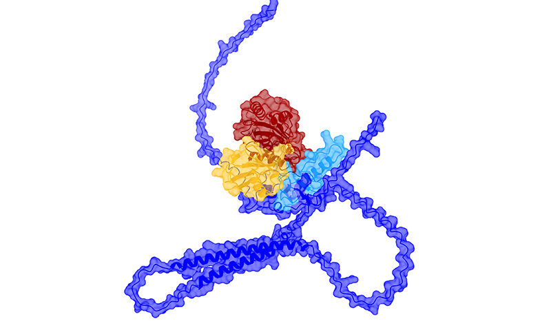
could be viewed in mammary ducts (blue) using the
new imaging technique. Only a few of these
pre-cancerous clones developed further to form a tumour.
Image from Rios, Visvader et al, Cancer Cell.
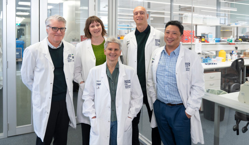
Imaging technology enabled the research team to examine the frequency of pre-cancerous cells that develop into tumours in the mammary gland, and address the behaviour of cells within tumours. The study was led by Dr Anne Rios with Professor Jane Visvader and Professor Geoff Lindeman at the Walter and Eliza Hall Institute.
“Using a new imaging technique, we revealed that only a small proportion of pre-cancerous cells will develop into tumours,” Professor Visvader said. “In contrast, once a tumour has been formed, we discovered it was very likely for its cells to undergo a so-called ‘epithelial-to-mesenchymal transition’ (EMT). This is a change in the ‘molecular landscape’ – the genes that are switched on or off – within the cell, transforming it from an ‘epithelial’ form, to a ‘mesenchymal’ form that could have a growth advantage.
“Our models suggest that EMT is not a rare event but is an inherent feature of mammary tumour cells.”
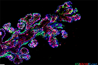
allows researchers to see into tissues at high resolution.
Image from Rios, Visvader et al, Cancer Cell.
The results were obtained in laboratory models that closely resemble human breast cancer, and the team suspects human breast cancers are likely to also show a high rate of molecular EMT, said Professor Lindeman, who is also a medical oncologist at the Royal Melbourne Hospital and the Peter MacCallum Cancer Centre.
“If EMT frequently occurs in breast cancers, it means the cells are a ‘moving target’ – they can evade one set of weapons we have to fight the cancer, meaning we need to develop strategies that are more broadly targeted,” said Professor Lindeman.
New views into tumours
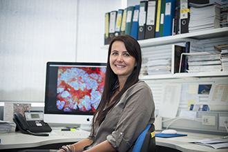
Hall Institute
A new three-dimensional imaging technique was critical for the discoveries said Dr Rios, who now works at the Princess Máxima Center for Pediatric Oncology in the Netherlands.
“Until now it has been challenging to visualise the intricate structures of complex tissues such as breast tissue, or to see the true arrangement of cells within tumours,” Dr Rios said. “We developed a new, rapid way to prepare tissue samples that retains their intricate architecture but allows us to distinguish individual cells and the three-dimensional structure of the tissue.
of mammary ducts in human breast tissue.
Image from Rios, Visvader et al, Cancer Cell.
“Our method enabled us to capture previously unseen images of breast tissue and mammary tumours – this was crucial for us to discover the frequency of EMT within the tumours.
“We expect there will be many other applications for our new imaging method, to study normal and cancerous tissue samples,” Dr Rios said.
The research also involved collaboration with the Institute’s Centre for Dynamic Imaging and Bioinformatics division.
The research was supported by funding from the Australian National Health and Medical Research Council, the National Breast Cancer Foundation, Cure Cancer Australia, the Australian Cancer Research Foundation and the Victorian Government.
Three-dimensional view of the structure of a mammary duct. Video from Rios, Visvader et al, Cancer Cell.
Media inquiries
M: +61 475 751 811
E: communityrelations@wehi.edu.au
Super Content:
Want to hear about our latest discoveries? Subscribe to our supporter newsletter, Illuminate.
This animation from WEHI.TV visualises research published in Nature Medicine in 2009 by Professor Jane Visvader and Professor Geoff Lindeman.
Video 1:06
Optical microscopy has become one of the most powerful tools in medical research.
WEHI's Centre for Dynamic Imaging is advancing our understanding of how diseases develop, spread and respond to treatment.
Professor Geoff Lindeman discusses his team's findings