Iris: Nanoflower Unfolding
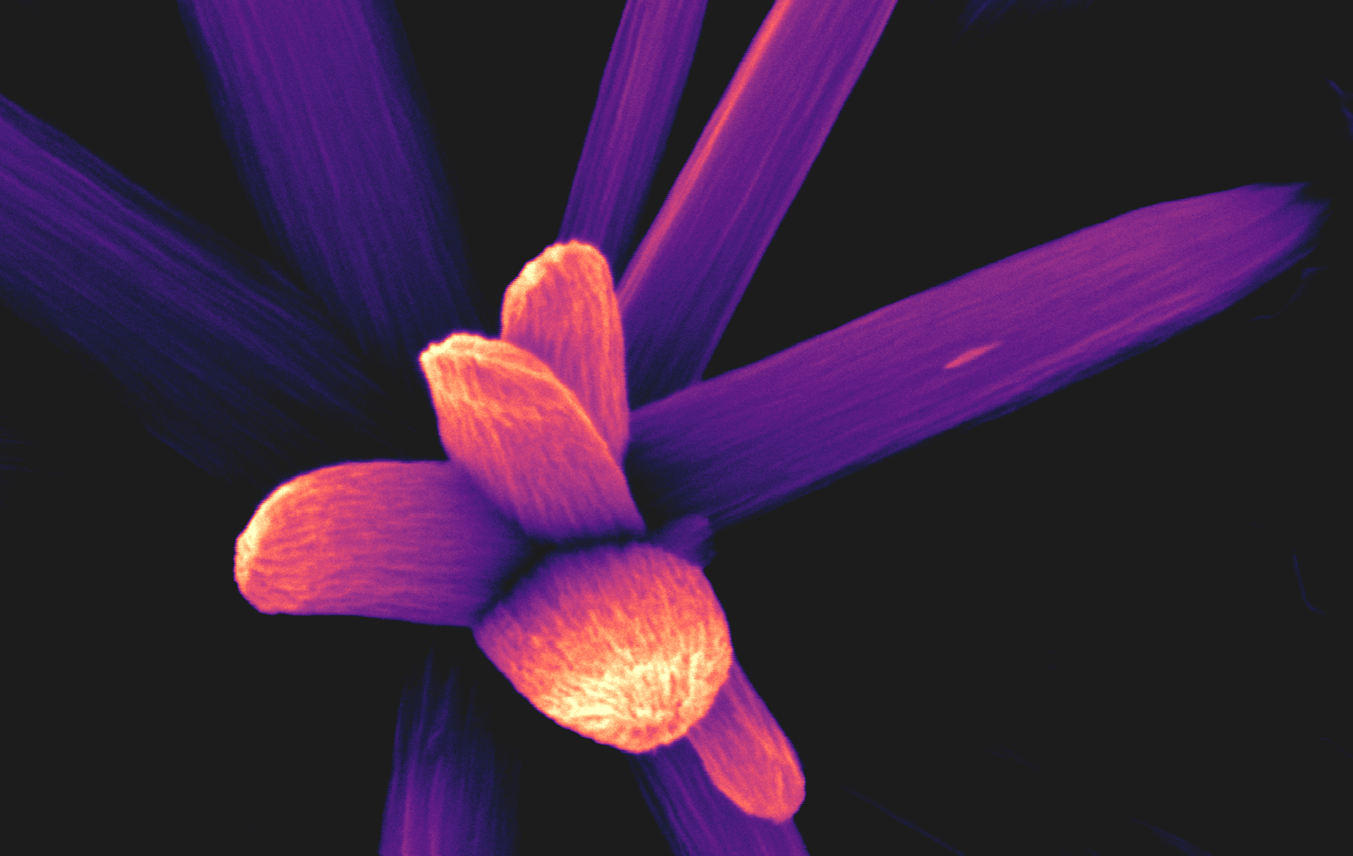
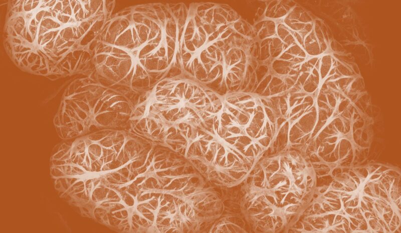
This impression of an iris in bloom is an image of tiny copper wires, called nanowires, captured by an electron microscope. These nanowires are grown via corrosion on a specimen support grid. They are used to examine protein molecules and help researchers identify linkages to disease.
The nanowires help spread a protein solution, creating thin samples essential for high-resolution electron microscopy. The solution is applied to the grid, flash frozen, then photographed at low temperatures – revealing intricate details.
Like unfolding the petals of a budding flower, such investigations reveal the inner ‘molecular machines’ that can drive disease. Studies like these inform the design of cancer drugs aiming to have fewer side effects than traditional chemotherapy or radiotherapy.
Artists
- Andrew Leis
- Mihin Perera
- Shabih Shakeel