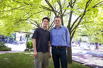
(L-R) Dr Wilson Wong, Professor Alan Cowman and collaborators
have visualised a key molecular structure that could help to design a
vaccine against the deadliest malaria parasite, Plasmodium falciparum.
An international team led by Institute researchers has visualised the unique molecular ‘key’ used by the world’s deadliest malaria parasite, Plasmodium falciparum, to enter and infect human blood cells.
This breakthrough provides scientists with the missing information required to design a vaccine that combats the prevalent parasite.
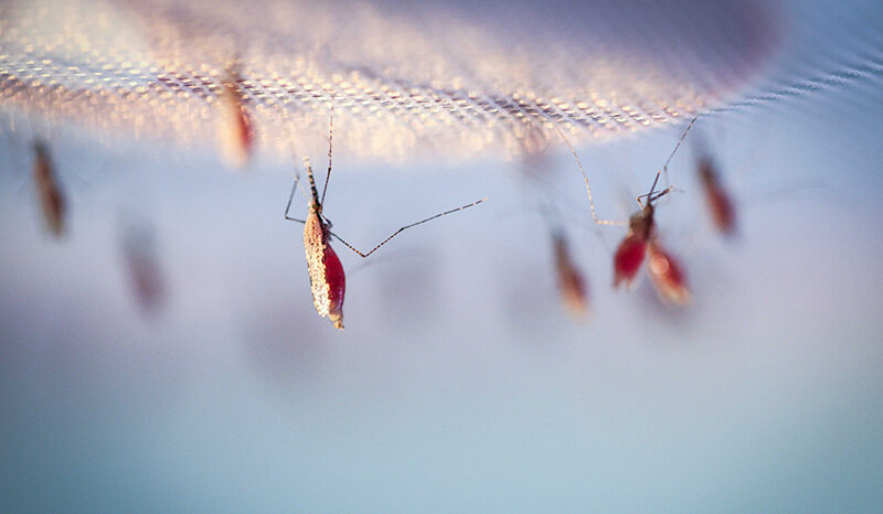
The findings represent a significant milestone because the malaria parasite kills more than 500,000 people each year and an effective vaccine for protection against it does not yet exist.
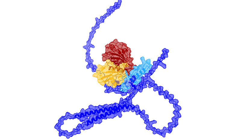
The unique molecular ‘key’ used by the world’s deadliest malaria parasite to enter human blood cells has been captured for the first time.
Cryo-electron microscopy enabled the researches to produce a detailed 3D ‘blueprint’ of the proteins that come together to form this special key.
The information can now be used to block the parasite from entering blood cells which means it will be unable to cause malaria infection in the body.
The ‘key’ to infection
The first-ever 3D image of the parasite’s key to causing infection was achieved using Nobel Prize-winning cryo-EM (cryo-electron microscopy) technology.
The ‘key’ is a complex of three parasite proteins – called Rh5, CyRPA and Ripr – which work together to unlock and enter the cell, Professor Cowman said.
“This complex is fundamental to the malaria parasite’s ability to enter cells and cause infection. With this new information we can now target the parasite in a much better way because we understand how it functions to infect the blood.
“Capturing the first ever image of the protein complex – revealing with astounding clarity exactly what it looks like – was a ‘Eureka’ moment in the field of malaria research,” he said.
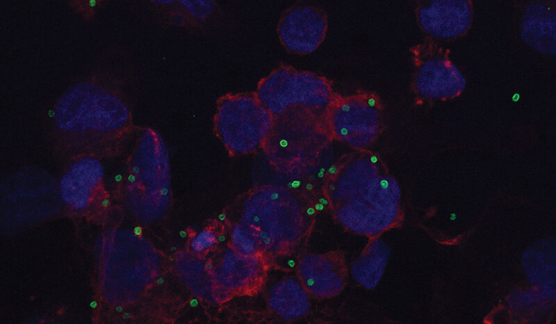
These findings are significant because the entry of the malaria parasite into human red blood cells enables rapid growth, multiplication and spread; driving serious symptoms such as fever, chills, malaise, diarrhoea and vomiting. Understanding how parasites enter cells opens up opportunities for blocking the parasite from infecting humans and stopping the cycle of disease and transmission.
Seen for the first time
The Melbourne-based team led by Professor Cowman prepared samples for the study by genetically engineering parasite DNA and extracting the proteins Rh5 and CyRPA. The third protein, Ripr, was produced by the biotechnology company ExpreS2ion.
Dr Wong said the 3D image of the ‘Rh5/CyRPA/Ripr’ complex was obtained using the world’s most advanced cryo-electron microscope, the Titan Krios, at the Howard Hughes Medical Institute’s Janelia Research Campus.
“Together with our colleagues in the US we obtained hundreds of thousands of images of the complex from different angles.
“With the help of high-powered computing we were then able to assemble these together, revealing the first-ever high resolution, 3D image of the Rh5/CyRPA/Ripr complex – the key to the parasite’s ability to cause infection,” Dr Wong said.
‘Blueprint’ makes vaccine possible
Professor Cowman said the new structure provided researchers with critical information for designing an effective vaccine against the Plasmodium falciparum parasite.
“We now have the information required to design a vaccine that gives the immune system precise instructions about how to stop the malaria parasite.
“If we can block the protein complex from forming, Plasmodium falciparum will never have the key it needs to infect human blood cells,” he said.
“Making this discovery has been rewarding because it brings us an important step closer to hopefully one day achieving the ultimate goal of eradicating malaria.”
The work builds on foundational studies led by Walter and Eliza Hall Institute researchers, to understand the role of Rh5, CyRPA and Ripr in malaria infection.
The research was supported by the Australian National Health and Medical Research Council and the Victorian Government.