A focus on world-leading microscopy

A focus on world-leading microscopy

Illuminate newsletter header, Autumn 22
March 2022
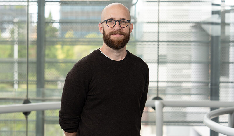
As a school student in the United States, Dr Michael Mlodzianoski had a natural ability for physics and mathematics. By the time Michael was at university, his timetable reflected a deep passion for science.

Dr Michael Mlodzianoski
Dr Michael Mlodzianoski designs and builds custom
microscopes for the WEHI Centre for Dynamic Imaging facility.

During his Master’s degree at the University of Maine, USA, Michael worked under a new professor who was establishing a microscopy and biophysics lab. The experience was pivotal for Michael – it was microscopy that he wanted to focus his career on.

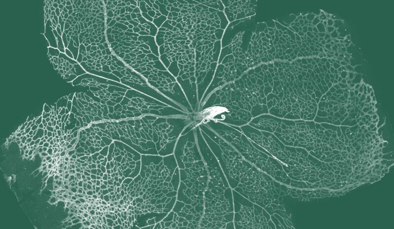
Fast forward a few years and a change of continents. These days you will find Michael in WEHI’s Centre for Dynamic Imaging where he is instrumental in helping scientists use advanced imaging technology and powerful equipment to create detailed, real-time views of biological systems.

“I’m learning more than I could have ever imagined about the biggest disease challenges of our time,” he said.

Michael is currently working to design and build microscopes to address the needs of WEHI researchers and the health problems they are working to solve.

“Establishing a foundation to build custom microscopes means we can quickly respond to new and exciting developments in microscopy,” he said. “This is vital for remaining at the forefront of research across several fields, whether it’s trying to see how cancers spread, or how our immune cells work to protect us.”

The crucial insights gained are advancing our understanding of how diseases develop, spread and respond to treatment.

Super Content: 
Lattice light sheet microscope

Optical microscopy has become one of the most powerful tools in medical research.

WEHI's Centre for Dynamic Imaging is advancing our understanding of how diseases develop, spread and respond to treatment.

Four researchers standing in the WEHI garden

A new imaging technique is shining a light on immune responses and setting the scene for enhancing immune memory to optimise vaccine strategies.

Microscopy image of cancer cells

Single cell imaging techniques are allowing researchers to develop complex mathematical models to understand how cancer cells respond to chemotherapy.