
Aurora
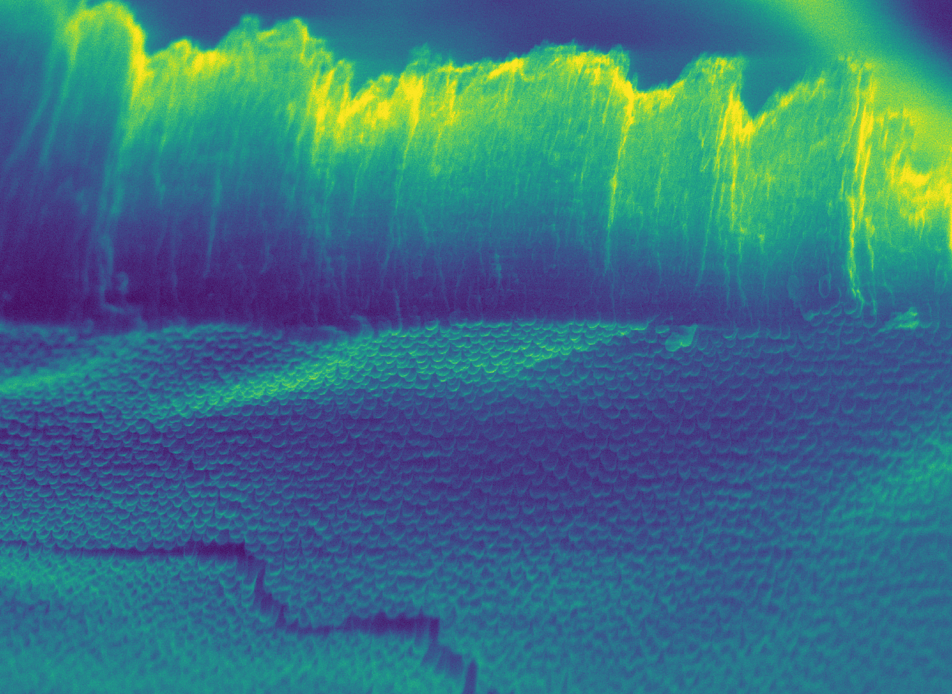
Resembling the northern lights, this image shows the underside of a coating developed for a nanofabricated microscope slide. WEHI and University of Melbourne researchers adapted the slide to study proteins through 3D cryo-electron microscopy.
The aurora waves are the cross section of the coating while the blue and green ‘treetops’ at the bottom of the image are closed ends of tiny holes, each 1000 times thinner than a human hair.
Researchers add droplets of protein molecules onto these surfaces, which are frozen and transferred into an electron microscope. The molecules are photographed at about 100,000 times magnification to visualise their structure.
Knowing the structure of a disease-causing protein can help scientists develop more precise treatments that target the right molecule, potentially increasing effectiveness and reducing side-effects for patients.
- Mihin Perera
- Andrew Leis
- Shabih Shakeel
- Maryam Zaman
- Joseph Berry (University of Melbourne)