An international study co-led by Institute researchers could help to make therapeutic insulins more effective by better mimicking the way insulin works in the body. The findings could improve treatments for diabetes, a disease that impacts the lives of millions of people worldwide.
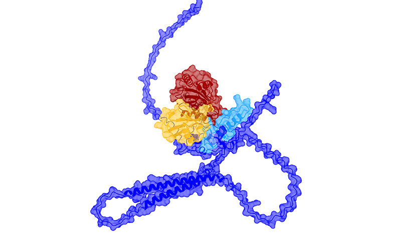
Published today in Nature Communications, the study reveals the first definitive 3D image of how insulin successfully interacts with its receptor — a ‘gatekeeper’ for transmitting information into cells— in a process that is crucial for lowering blood sugar levels in the body. Understanding exactly what this process looks like can inform the design of faster-acting and longer-lasting insulin therapies.
The research was co-led by Associate Professor Mike Lawrence from the Walter and Eliza Hall Institute, in collaboration with teams led by Dr Matthias Dreyer from the pharmaceutical company Sanofi-Aventis Deutschland GmbH and Dr Christoph Müller at the European Molecular Biology Laboratory (EMBL), both based in Germany.
At a glance
An international collaboration co-led by Institute researchers has revealed how insulin triggers cells to reduce blood sugar levels in the body.
The researchers have produced the first 3D image of how insulin binds to its receptor on the surface of cells and transmits into the cell instructions to take up sugar from the blood.
This image is a game-changer for designing both faster-acting and longer-lasting insulin therapies that will help to reduce the burden of diabetes worldwide.
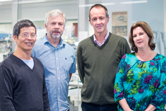
(L-R) Dr Yibin Xu, Dr John Menting, Associate Professor Mike Lawrence and Mai Margetts.
The missing piece of the puzzle
Associate Professor Lawrence said he was thrilled to have resolved the long-standing question of how insulin binds to its receptor and triggers cells to lower blood sugar levels.
It is well established that insulin instructs cells to lower blood sugar levels in the body by binding to a receptor that is located on the cell surface.
Associate Professor Lawrence said the problem was no one knew precisely what was occurring during the interaction.
“Current insulin therapies are sub-optimal because they have been designed without this crucial knowledge,” he said.
“Together with our collaborators in Germany, we have found that missing piece of the puzzle in producing the first definitive 3D image of the way in which insulin binds to the surface of cells in order to successfully transmit the vital instructions needed for taking up sugar from the blood.”
Seeing is believing
Associate Professor Lawrence said the detailed image was the outcome of a collaboration between structural and cell biology experts from the Institute working together with both cryo-electron microscopy specialists at EMBL in Heidelberg and an insulin receptor specialist from the University of Chicago.
“We knew that insulin underwent a physical change that signalled its successful connection with its receptor on the cell surface. But what we had not seen before were the detailed changes that occurred in the receptor itself, confirming that insulin had successfully delivered the message for the cell to take up sugar from the blood.
“My colleagues at the Institute carefully engineered individual samples of insulin bound to receptors so that our collaborators in Heidelberg could use cryo-electron microscopy to capture hundreds of thousands of high-resolution ‘snap shots’ of these samples.
“We then combined more than 700,000 of these 2D images into a high-resolution 3D image showing precisely what the successful binding between insulin and its receptor looks like. And there it was before our eyes, the full picture in exquisite detail.
“It was at that point we knew that we had the information needed to develop improved insulin therapies that could ensure cells would respond correctly and carry out the functions necessary to lower blood sugar levels,” Associate Professor Lawrence said.
Optimal therapies now possible
Associate Professor Lawrence said the findings meant it would now be possible to design insulin therapies that could mimic more closely the body’s own insulin.
“Going forward, pharmaceutical companies will be able to use our data as a ‘blueprint’ for designing therapies that optimise the body’s uptake of insulin. There has already been great interest in these results and their application, and the Institute has a network of collaborations already underway.
“After more than two decades of research by Melbourne scientists to get to this point, it is phenomenal to have achieved results that will ultimately benefit patients with the development of more effective therapies, particularly for those whose lives are dependent on a daily injection of insulin,” he said.
The research was funded in part by the Australian National Health and Medical Research Council, the Victorian State Government and the Australian Government’s NHMRC Independent Research Institute Infrastructure Support Scheme.