$20M microscopy facility to help unlock secrets of diabetes, malaria
02 February 2015
Melbourne scientists now have access to an unparalleled view of the molecular world thanks to Australia’s first advanced structural cryo-electron microscopy centre.
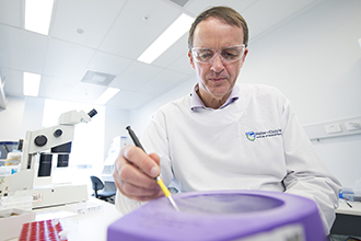
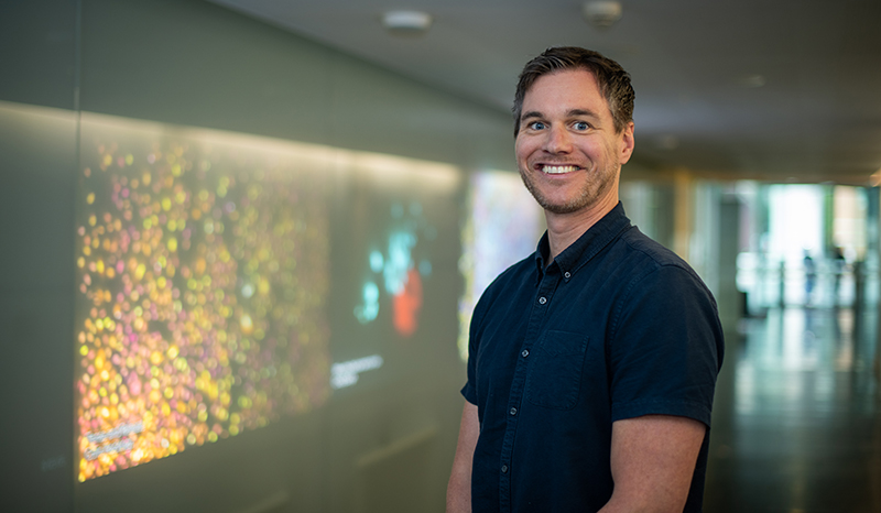
Associate Professor Mike Lawrence said the cryo-electron microscope would aid his diabetes research.
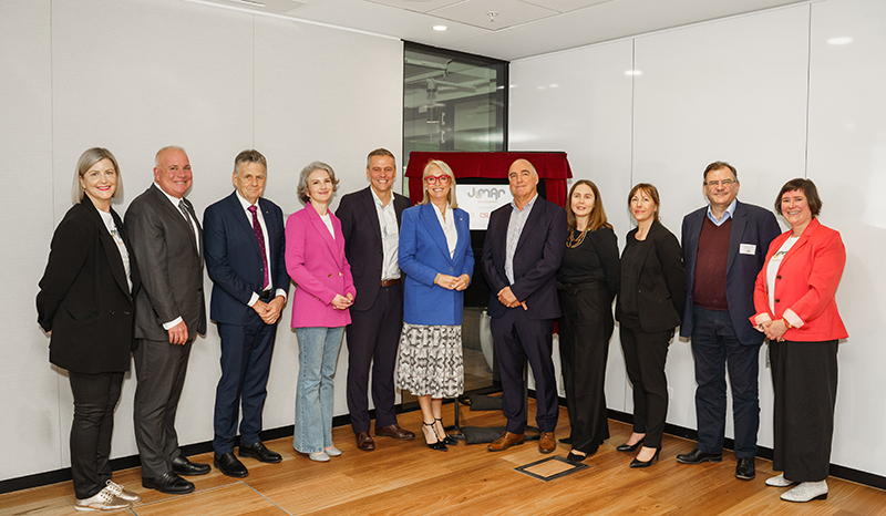
The $20 million Clive and Vera Ramaciotti Centre for Structural Cryo-Electron Microscopy was launched today at Monash University, Clayton. Professor James Whisstock from Monash University will be the acting director, with Associate Professor Mike Lawrence from the Walter and Eliza Hall Institute coordinating the institute’s role as a foundation partner in the centre.
The centre features a $5 million Titan Krios cryo-electron microscope, which is powerful enough to resolve three-dimensional molecular shapes and structures with detailed precision, creating exquisite models of important biological molecules.
Investigating insulin’s action
Associate Professor Lawrence said visualising how insulin bound to its receptor was crucial to understanding how diabetes developed. “Type 2 diabetes is characterised by reduced sensitivity to insulin, and discovering how this hormone interacts with the receptor could shed light on the mechanisms behind this disease,” he said.
“The structure of insulin has been known for many decades, however we have only recently determined how insulin binds to its receptor. Previous imaging techniques have limited our ability to visualise how this interaction occurs as we could only see fragments of the receptor at a time. This new technology will enable us to create three-dimensional structures of the whole insulin receptor, vastly improving our understanding of this critical molecule.”
Tackling malaria
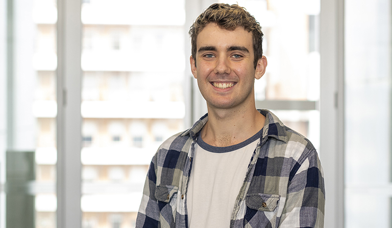
Dr Wilson Wong recently travelled to the UK to use a Titan Krios cryo-electron microscope to study the ‘protein factory’ of the malaria parasite.
Having this technology available in Australia would enable researchers to visualise biological structures without the need for crystallisation and with only relatively small quantity of materials, Dr Wong said. “The best imaging technologies available, such as X-ray crystallography, requires researchers to crystallise the proteins they are investigating,” he said. “This process can take days, weeks or even months, and is not always successful.
“Using this microscope will mean scientists can obtain high-resolution images of large protein complexes that are difficult or impossible to crystallise.”
Dr Wong said he would use the new facility to continue investigating other parts of the molecular machinery critical for the malaria parasite’s survival. “We hope to identify potential drug targets that could be used to treat or eradicate this disease,” he said.
The Clive and Vera Ramaciotti Centre for Structural Cryo-Electron Microscopy is jointly funded by the Ramaciotti Foundations, Australian Research Council, Monash University, Walter and Eliza Hall Institute, La Trobe University and the National Health and Medical Research Council.